

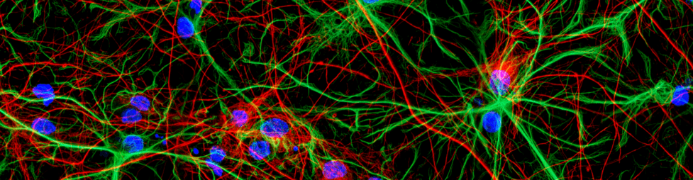
Growth of Cerebral Organoids
Welcome to this guide on generating cerebral organoids starting from H9 embryonic stem (ES) cells, adapted from the methods established by Lancaster and colleagues. In this approach, ES cells or induced pluripotent stem cells (iPSCs) are first separated into single cells from their feeder layers. They are then expanded in 96-well plates to form embryoid bodies, before moving through a series of media changes to induce neural fate and eventually promote 3D differentiation in a spinning bioreactor. Let's dive into the details to help you get your 3D brain models up and running!
Featured Key Products
TCB-32 (Small Molecule FGF2 Replacement)
Synthetic, thermostable FGF2 replacement. Suitable for cell culture.
View ProductTCB-541 (Small Molecule FGF2 Replacement)
Highly potent synthetic, thermostable FGF2 replacement. Suitable for cell culture.
View ProductTCB-621 (Small Molecule FGF2 Replacement)
Highly potent synthetic, thermostable FGF2 replacement. Suitable for cell culture.
View ProductROCK inhibitor Y-27632
Selective ROCK inhibitor frequently used for the production of organoids
View ProductMaterials
- H9 ES cells or iPSCs
- Basement membrane extract (generic alternative)
- FGF2 or TCB Small Molecule FGF2 Replacements
- ROCK inhibitor Y-27632 (HB2297)
- DMEM:F12 medium
- Neurobasal medium
- N2 supplement
- B27 supplement (with and without vitamin A)
- Glutamax or equivalent L-glutamine supplement
- 2-Mercaptoethanol
- MEM-NEAA
- Insulin
- Heparin sodium salt
Media Recipes
Aseptically combine components and filter the media before use.
hES Media
| Component | Concentration | |
|---|---|---|
| ROCK inhibitor Y-27632 (HB2297) | 50 μM | |
|
One of: |
FGF2 |
4 ng/ml |
| TCB-32 (HB12632) | 400nM | |
| TCB-541 (HB11050) | 200nM | |
| TCB-621 (HB17550) | 100nM | |
Please note: FGF2 can be replaced with stable, cost-effective small molecule FGFR1 agonists TCB-32, TCB-541, or TCB-621 to enable weekend-free feeding and significantly reduce media costs. TCB compounds have not been optimised yet for this assay therefore concentrations may need adjusting. TCB concentrations are based upon the original 5x lower FGF2 concentration used by Lancaster et al..
Neural Induction Media
| Component | Concentration |
|---|---|
| DMEM:F12 medium | 100% |
| N2 Supplement | 1:100 |
| Glutamax | 1:100 |
| MEM-NEAA | 1:100 |
| Heparin sodium salt | 1 μg/ml |